How is Early Evaluation Done During Pregnancy?

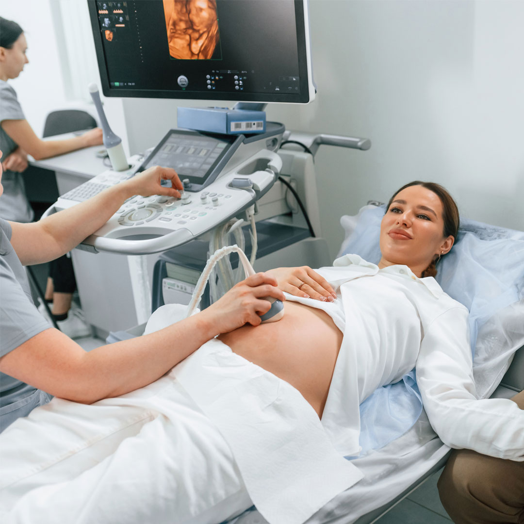
Evaluation of fetal anomalies and genetic anomalies during pregnancy is a complicated process. Evaluations that begin even before pregnancy are important. Especially the first trimester (first 12 weeks of pregnancy) evaluation is very important for early diagnosis. Structures that need to be examined with ultrasonographic evaluation this week:
- fetal head
- limbs
- abdominal structure
- structure of the heart
- Tricuspid valve flow
- nasal bone
- nuchal translucency
- Intracranial translucency
- Ductus venous flow
- Placenta
- Cervical length, etc.
Defects in these structures are important for early diagnosis. Early recognition provides the chance of earlier intervention. The importance of genetic examination increases in the presence of certain findings on ultrasound. Increase in neck thickness, smallness of the nasal bone, deviations from normal blood flows are warning signs of genetic diseases.
Even if no problem is detected in the ultrasound examination, it does not mean that the baby will not have a genetic disease. For genetic examination, the family is discussed and the examination is carried out using whatever method the family wishes. There are several methods for genetic analysis. First of all, the presence of normal nuchal thickness and nasal bone reduces the possibility of genetic diseases. If the family does not want to be examined with another method, the family is explained that this method has a low chance of success and follow-up continues. A superior method of examination is double testing. Ultrasound findings are combined with the blood sample taken from the mother to produce a statistical result. According to this result, the possibility of a genetic disease is explained to the family. Although the chance of catching a genetic disease is higher than the prediction made based on nuchal thickness alone, the success rate of catching genetic diseases is not very high.
Today, another method with a higher success rate in making a diagnosis is the NIPT (non-invasive prenatal diagnosis) method. It is the examination of the baby's DNA in the mother's blood. The success rate is higher than the double test. All of these methods are frequently used in clinical practice, but none of them are diagnostic methods. It is considered a screening test and gives the numerical value of the probability of having a genetic disease in the baby. The diagnostic method is CVS (chorionic villus sampling), which we perform when necessary. In this procedure, similar to amniocentesis, a very thin needle is taken from the structure called placenta, from which the baby is fed. It is a diagnostic method.
As a result, all pregnant women should undergo a perinatological examination in the first weeks of their pregnancy.
Dr. Baris Sever
Gynecology and Obstetrics
Perinatology Specialist
Contact Us For Appointment:
Telephone line: 0392 444 3548 (ELIT)
Contact Form: https://www.elitenicosia.com/iletisim/


