MRI-TRUS Prostate Fusion Biopsy Procedure: Current Diagnostic Approach
Entrance
Prostate cancer is one of the most common malignancies in men, and early diagnosis and accurate staging directly affect treatment success.
Currently, standard transrectal ultrasound (TRUS)-guided biopsies may be inadequate in detecting clinically significant prostate cancer due to limited accuracy.
At this point, the MRI-TRUS fusion biopsy method is a modern diagnostic method that stands out thanks to its imaging and targeting advantages.
What is MRI-TRUS Fusion Biopsy?
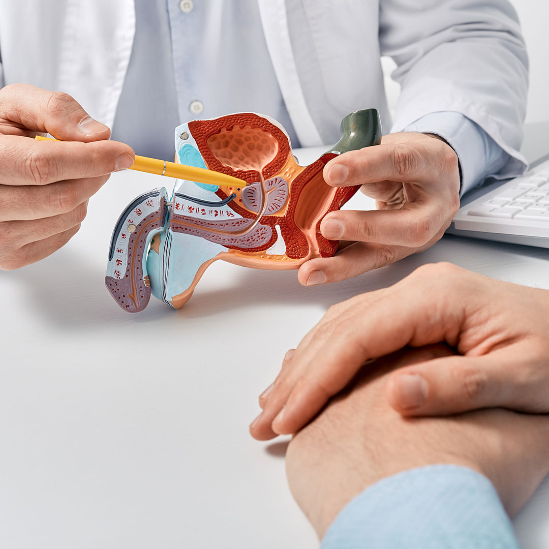
MRI-TRUS fusion biopsy is based on the principle of combining (fusion) high-resolution prostate images obtained by magnetic resonance imaging (MRI) with images taken in real time with transrectal ultrasound (TRUS) with the help of software.
MRI identifies suspicious foci in the prostate tissue.
TRUS provides live guidance during biopsy.
Thanks to the fusion software, suspicious areas are reflected on the ultrasound screen and the needle biopsy is directed to the target.
Implementation of the Process:
- MRI stageThe patient first undergoes a multiparameter prostate MRI. Suspicious lesions are classified using the PI-RADS (Prostate Imaging Reporting and Data System) scoring system.
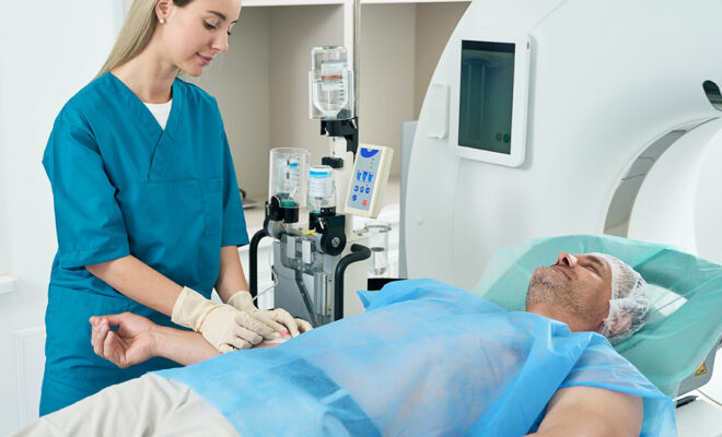
- TRUS stageOn the day of the procedure, the ultrasound probe is placed in the rectum under local or general anesthesia.
- Fusion: Special software matches previously acquired MRI images with TRUS.
- BiopsyTargeted sampling is performed on suspicious areas. Systematic biopsy may be added if necessary.
Advantages:
- Higher diagnostic accuracy: The rate of detecting clinically significant prostate cancer increases.
- Fewer unnecessary biopsies: Detection of insignificant or low-grade cancers is reduced.
- Targeted sampling: Unnecessarily many injection attempts are not made.
- Ease of treatment planning: The localization of the detected lesion is determined more clearly.
Post-Processing Process:
After the biopsy, patients may experience mild rectal discomfort and blood in the urine or semen. These findings usually disappear within a few days.
Prophylactic antibiotics are used due to the risk of infection. The rate of serious complications is quite low.
Conclusion:
MRI-TRUS fusion biopsy is considered a superior method compared to classical TRUS biopsies in the diagnosis of prostate cancer.
It is especially preferred in patients with elevated PSA levels but negative results in previous biopsies or in whom a suspicious focus is detected on MRI.
With developing technology, this method is expected to become the standard approach in the diagnosis of prostate cancer.
Specialist Dr. Mehmet ADIGÜZEL
Radiology Specialist – Elite Hospital